
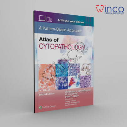

Atlas of Cytopathology: A Pattern-Based Approach
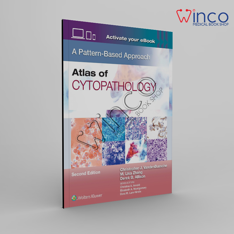
Atlas of Cytopathology: A Pattern-Based Approach
Shipping: Delivery 2 To 3 Working Days
Returns: 7 Days Easy Return
ORDER ON WHATSAPP | واٹسّپ پر آرڈر کریں

Atlas of Cytopathology: A Pattern-Based Approach – Comprehensive
Atlas of Cytopathology: A Pattern-Based Approach is a comprehensive educational reference that provides a practical, visually rich, and systematic guide to the interpretation of cytopathology specimens. Designed for pathologists, pathology residents, cytotechnologists, laboratory professionals, medical students, and clinicians, this atlas emphasizes a pattern-based diagnostic strategy that simplifies complex cytological findings into logical, reproducible diagnostic pathways. By integrating high-quality photomicrographs, detailed explanations, differential diagnoses, and evidence-based diagnostic principles, the book serves as an invaluable resource for both everyday clinical practice and examination preparation.
Unlike traditional cytopathology textbooks that primarily organize content according to organ systems or disease categories, this atlas adopts a pattern-recognition methodology. Readers first learn to identify characteristic cellular patterns before progressing toward specific diagnoses. This practical approach mirrors the real-life workflow used by experienced cytopathologists and significantly enhances diagnostic confidence, particularly when evaluating fine-needle aspiration (FNA) specimens, exfoliative cytology, and body fluid samples.
The atlas begins with a strong foundation in the principles of diagnostic cytopathology. Readers are introduced to specimen collection techniques, smear preparation, liquid-based cytology, fixation methods, staining procedures, slide quality assessment, laboratory safety, quality assurance, and specimen adequacy criteria. These introductory chapters establish the essential knowledge required to understand cytological interpretation while emphasizing best laboratory practices that ensure accurate and reliable diagnoses.
A major strength of this book is its exceptional collection of high-resolution microscopic images. Every chapter contains carefully selected photomicrographs demonstrating normal cellular morphology, benign reactive changes, inflammatory processes, premalignant lesions, and malignant neoplasms. Multiple examples illustrate the spectrum of morphologic variation seen in routine diagnostic practice, enabling readers to recognize subtle differences that distinguish benign conditions from clinically significant pathology.
The pattern-based framework encourages readers to analyze specimens according to observable cytological features rather than memorizing isolated diseases. Nuclear enlargement, chromatin characteristics, nucleolar prominence, cytoplasmic appearance, architectural arrangements, background material, inflammatory components, and extracellular matrix patterns are systematically examined to guide accurate differential diagnosis. This structured methodology improves consistency while reducing diagnostic errors.
Comprehensive coverage of fine-needle aspiration cytology forms one of the central components of the atlas. Detailed discussions explain aspiration techniques, specimen handling, rapid on-site evaluation (ROSE), cell block preparation, ancillary testing, and interpretation of aspirates obtained from various anatomical sites. Readers learn how to correlate clinical information, imaging findings, and microscopic morphology to produce meaningful diagnostic reports that directly influence patient management.
The thyroid chapter provides an in-depth review of thyroid cytopathology using contemporary reporting systems and diagnostic terminology. Normal thyroid tissue, colloid nodules, Hashimoto thyroiditis, Graves disease, follicular neoplasms, papillary thyroid carcinoma, medullary carcinoma, anaplastic carcinoma, and metastatic lesions are illustrated with abundant cytologic images. Characteristic nuclear grooves, intranuclear inclusions, colloid quality, architectural patterns, and diagnostic pitfalls are clearly explained to facilitate confident interpretation of thyroid FNA specimens.
Breast cytopathology is covered extensively through discussions of benign proliferative lesions, fibrocystic changes, fibroadenoma, inflammatory disorders, papillomas, atypical proliferative lesions, ductal carcinoma, lobular carcinoma, mucinous carcinoma, medullary carcinoma, and metastatic tumors. The atlas highlights cytological criteria used to distinguish benign epithelial proliferations from malignant lesions while emphasizing clinicopathologic correlation and appropriate ancillary studies.
The lymph node chapter introduces reactive lymphoid hyperplasia, granulomatous inflammation, infectious diseases, Hodgkin lymphoma, non-Hodgkin lymphoma, metastatic carcinoma, and hematologic malignancies. Readers become familiar with lymphoid cell populations, cytomorphologic features of various lymphoma subtypes, and the role of immunocytochemistry, flow cytometry, molecular diagnostics, and cytogenetic studies in establishing definitive diagnoses.
Salivary gland cytopathology is presented through an organized review of inflammatory lesions, benign neoplasms, and malignant tumors. Pleomorphic adenoma, Warthin tumor, mucoepidermoid carcinoma, acinic cell carcinoma, adenoid cystic carcinoma, and secretory carcinoma are described using distinctive cytological patterns supported by high-quality microscopic illustrations. Diagnostic challenges, overlapping morphologic features, and common pitfalls receive special attention.
Respiratory cytology chapters examine sputum cytology, bronchial brushings, bronchoalveolar lavage specimens, endobronchial ultrasound-guided aspirations, and transthoracic FNA samples. Benign respiratory changes, infections, inflammatory conditions, primary lung carcinomas, neuroendocrine tumors, metastatic malignancies, and mesothelioma are systematically reviewed. The atlas emphasizes cellular morphology, tumor classification, ancillary testing, and molecular biomarkers increasingly used in modern thoracic oncology.
Gynecologic cytopathology receives comprehensive coverage through detailed explanations of cervical screening, the Bethesda System, human papillomavirus-associated lesions, squamous intraepithelial lesions, glandular abnormalities, adenocarcinoma, endometrial pathology, vaginal cytology, and post-treatment surveillance. Numerous photomicrographs demonstrate both classic and uncommon cytological findings, allowing readers to confidently interpret cervical specimens while recognizing diagnostic mimics.
Urinary tract cytology chapters discuss urine specimens, bladder washings, upper urinary tract cytology, reactive atypia, instrumentation-related changes, infections, polyomavirus infection, urothelial carcinoma, carcinoma in situ, high-grade malignancies, and secondary metastatic involvement. Particular emphasis is placed on the Paris System for Reporting Urinary Cytology, improving diagnostic standardization and communication among healthcare providers.
The gastrointestinal and pancreaticobiliary sections describe cytological evaluation of esophageal lesions, gastric pathology, pancreatic masses, liver lesions, biliary strictures, and gastrointestinal stromal tumors. Endoscopic ultrasound-guided fine-needle aspiration is thoroughly discussed alongside cytomorphologic interpretation, specimen adequacy assessment, and the role of ancillary molecular testing in precision medicine.
The atlas also includes dedicated chapters covering liver cytology, soft tissue tumors, bone lesions, skin tumors, pediatric neoplasms, endocrine organs, and uncommon metastatic malignancies. These sections broaden the diagnostic scope while introducing readers to rare entities encountered in specialized pathology practice.
Body fluid cytology represents another major focus of the book. Pleural, peritoneal, pericardial, cerebrospinal, and synovial fluid specimens are evaluated using a pattern-based framework that distinguishes reactive mesothelial proliferations from metastatic carcinoma, lymphoma, mesothelioma, and infectious processes. Readers learn to identify subtle morphologic clues supported by immunocytochemistry and ancillary laboratory techniques.
One of the most valuable features of the atlas is its detailed differential diagnosis tables. Similar lesions are compared side by side using key cytological characteristics, clinical presentation, immunophenotypic markers, molecular findings, and diagnostic pitfalls. These comparison tables allow readers to rapidly differentiate morphologically similar conditions while reinforcing critical diagnostic principles.
Ancillary diagnostic techniques receive comprehensive attention throughout the book. Immunocytochemistry, flow cytometry, fluorescence in situ hybridization (FISH), polymerase chain reaction (PCR), next-generation sequencing (NGS), molecular biomarkers, digital pathology, and artificial intelligence-assisted image analysis are integrated into appropriate clinical contexts. Readers gain an appreciation of how these technologies complement traditional morphology to improve diagnostic precision.
Reporting systems standardized by international professional organizations are incorporated wherever applicable. The Bethesda System for thyroid and cervical cytology, the Milan System for salivary gland cytology, the Paris System for urinary cytology, the Yokohama System for breast cytology, and other internationally accepted classification systems are presented with clear explanations and practical examples. These standardized reporting frameworks improve communication between pathologists and treating clinicians while supporting evidence-based patient management.
Clinical correlation is emphasized throughout every chapter. Cytological findings are interpreted alongside patient history, physical examination, laboratory investigations, radiologic imaging, and histopathologic confirmation whenever appropriate. This multidisciplinary perspective reflects modern diagnostic practice and strengthens the reader’s ability to integrate diverse clinical information into accurate diagnostic decision-making.
Educational features significantly enhance learning efficiency. High-yield diagnostic pearls summarize important concepts, while caution boxes highlight common diagnostic pitfalls and interpretive challenges. Flowcharts guide readers through complex differential diagnoses, and summary tables consolidate essential information into accessible reference formats suitable for rapid review before examinations or sign-out sessions.
Case-based learning forms another important component of the atlas. Realistic clinical scenarios demonstrate how cytological findings influence patient care from initial presentation through diagnosis, treatment planning, and follow-up. These case discussions encourage critical thinking while reinforcing the practical application of morphologic principles in routine pathology practice.
The atlas also addresses quality assurance, laboratory accreditation, specimen adequacy, error prevention, diagnostic reproducibility, and continuous quality improvement. Best practices for laboratory workflow, slide review, documentation, consultation, and interdisciplinary communication are explained in detail, supporting safe and reliable patient care.
Modern developments in digital pathology are thoughtfully integrated into the text. Whole-slide imaging, telecytology, remote consultation, computer-assisted diagnosis, artificial intelligence algorithms, and machine learning applications are introduced as emerging technologies that are transforming cytopathology practice worldwide. Although traditional microscopic interpretation remains fundamental, readers gain insight into how technological innovation is reshaping future diagnostic workflows.
For pathology trainees preparing for board examinations, the atlas serves as an outstanding review resource. The organized pattern-based format facilitates rapid revision while strengthening visual memory through repeated exposure to classic cytologic images. Examination-style summary points reinforce frequently tested concepts, helping learners build confidence for certification and licensing examinations.
Experienced practicing pathologists also benefit from the atlas as a dependable reference during challenging diagnostic cases. Comprehensive image collections, differential diagnosis tables, ancillary testing recommendations, and reporting guidelines provide immediate support when evaluating uncommon lesions or resolving difficult morphologic interpretations.
Medical students and clinicians with limited pathology experience will appreciate the book’s clear writing style and progressive organization. Complex cytological concepts are explained using straightforward language without sacrificing scientific accuracy. Step-by-step diagnostic reasoning makes the material accessible while maintaining sufficient depth for advanced learners.
The visual design of the atlas further enhances usability. Consistent chapter organization, color-coded headings, labeled photomicrographs, detailed figure legends, diagnostic algorithms, and concise summaries allow readers to navigate efficiently between topics. Whether used for systematic study or quick consultation during clinical practice, information is presented in a logical and user-friendly manner.
Ultimately, Atlas of Cytopathology: A Pattern-Based Approach combines outstanding microscopic imagery, evidence-based diagnostic principles, standardized reporting systems, and practical clinical guidance into a single authoritative resource. Its pattern-recognition philosophy mirrors real-world diagnostic thinking, enabling readers to interpret cytologic specimens with greater confidence, accuracy, and consistency.
Whether learning the fundamentals of cytopathology, preparing for examinations, improving diagnostic skills, teaching trainees, or managing complex clinical cases, this atlas provides an exceptional educational foundation. Through its emphasis on morphology, differential diagnosis, ancillary testing, quality assurance, and multidisciplinary clinical correlation, Atlas of Cytopathology: A Pattern-Based Approach remains an indispensable reference for anyone dedicated to excellence in diagnostic cytopathology.
WINCO ONLINE MEDICAL BOOKS SHOP
Related products
-17%
-17%
Emergency Imaging of At-Risk Patients: General Principles 1st Edition
Original price was: ₨ 1,195.₨ 995Current price is: ₨ 995.
-20%

Notes and Notes for MRCP 1 and 2 (3 Volumes)
Original price was: ₨ 4,995.₨ 3,995Current price is: ₨ 3,995.
-21%
Differential Diagnosis in Pediatrics 1st Edition
Original price was: ₨ 1,895.₨ 1,495Current price is: ₨ 1,495.
-18%
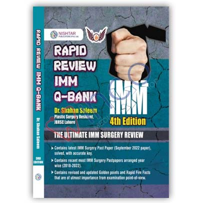
Rapid Review IMM Q Bank by Dr Shahan Saleem
Original price was: ₨ 1,095.₨ 895Current price is: ₨ 895.
-19%

Radiant Notes Fast Aid 5th Edition 2023 By Dr Rafi ULLAH
Original price was: ₨ 2,700.₨ 2,200Current price is: ₨ 2,200.
Select options
This product has multiple variants. The options may be chosen on the product page
-18%